Nuclear Medicine
Types of examinations and required preparations
| Examination | Preparation |
|---|---|
| Thyroid | None |
| Parathyroid | None |
| Salivary glands | None |
| Bone | Waiting 2 or 3 hours and drink 3 to 4 glasses of water between the 2 parts of the exam. |
| DMSA | None |
| DTPA | Keep well hydrated 01 hour before the examination |
| Lung | None |
| Abdomen | Fasting from midnight the day before the examination |
| MIBG | Lugol Preparation: drops solution to be taken 3 days before the examination and 2 days later to block the thyroid gland. |
| Iodine 131 Scan | None |
| Myocardium | To fast 4 hours before the examination. Avoid stimulating drinks and food (tea, coffee, chocolate ... etc.) 12 hours before the examination. Discontinuation of treatment accordi ng to cardiologist's instructions |
Contra-indications
Pregnancy.
If breastfeeding, suspend breastfeeding 24hour for all exams and 48 hours for myocardial scintigraphy (draw milk and reject it).
Conduct of Examination
It has two phases:
- Injection of an intravenous radiotracer
- During phase 2, the camera records images
The waiting time between these two phases depends on the examination time: a few minutes for the thyroid scintigraphy to 2 hours for bone scintigraphy or even 6 hours for myocardial scintigraphy.
The duration of the examination varies from 5 to 30 minutes maximum.
Treatment of hyperthyroidisms with iodine 131 (IRATHERAPY)
It is an effective treatment, easy to use and it is done on an ambulatory basis.
Indication
- BASEDOW disease
- Toxic nodule
- Toxic multiheteronodular goiter
Contraindications
- Pregnancy
- Breastfeeding woman: permanently stop breastfeeding after iodine administration
- Women of childbearing age: consider contraception for a duration stretching from 06 to 08 months
Equipment
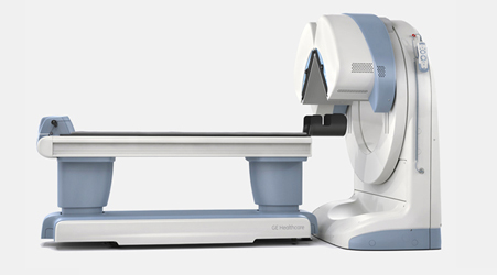
Device :02 double-Headed Gamma-Cameras with Xeleris 3 image processing unit
Supplier :General Electric
Type :Discovery NM630
Type :Discovery NM630

Technology
X-ray equipment provided with a digital plane sensor associated with a computer system. When X-rays are emitted, they pass through the structures of the examined area and will be intercepted by the plane sensor. The image is created by the computer conversion of the radiation into a multitude of points whose color goes from black to white depending on the intensity of the radiation.
The greater the amount of rays reaching the plane sensor, the more the created image will appear darker (the bone that blocks the rays significantly appears clear while the lungs that let many rays appear black).
What is the contribution of digital radiology ?
X-ray allows to analyze the possible modifications of a structure.
The contribution of scanning is important because it provides an editable image on a screen.
Opening hours
Saturday - Wednesday
07:00 - 16:00